Simple or complex, the microscope is the biologist’s most indispensable instrument. In an age when highly sophisticated and expensive instruments are the norm, professor of molecular and cellular biology Venkatesh N. Murthy wanted to encourage more of a “maker culture” among the department’s graduate students. So he taught them how to make one.
“For many years now in neuroscience,” Murthy explains, “advanced training has involved building things. There have been times when instruments were not easily available, and neuroscientists had to build their own equipment.”
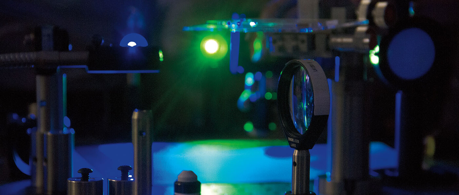
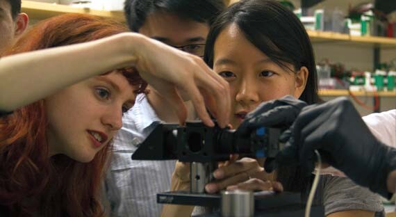
Murthy also wanted to deepen students’ knowledge of the fundamental principles of microscopy through experiential learning. “If you build something,” he says, “you’re going to understand it substantially more.” For the 20 participants in the class “Make Your Own Microscope,” that meant starting with “a bunch of parts,” including lenses, LEDs and lasers, a digital camera, and software to build a simple fluorescence microscope, the same off-the-shelf instrument used in biology labs, including Murthy’s, to study living cells.
Together with postdoctoral fellow Vikrant Kapoor, Murthy taught students how to use a light source to calculate the various properties of a lens, such as its focal length; how to use different light sources; how to assemble multiple lenses together to increase magnification; how to use filters to increase viewing contrast; and how to acquire images with a computer-controlled digital camera.
For Georgia Squyres, now a G2 in the Molecules, Cells, and Organisms (MCO) graduate program, learning to make a microscope was a chance to “get under the hood” of what at first glance appeared to be a very complex instrument. “As a biophysicist, I was excited to see an example of the very close relationship between engineering and life sciences,” Squyres explains. “I was able to experience, first hand, how a biological discovery—fluorescent proteins—promoted engineering advances—fluorescent microscopy—that let us ask novel biological questions.”
Squyres and her teammates started by “tinkering” to “understand the fundamentals,” and despite being “a little rough around the edges” they got it to work. Ultimately they captured images and videos of several biological samples, including the heartbeat of a zebrafish, which she says was “probably the most exciting because it’s dynamic.”
Now that’s she’s made a microscope, Squyres says she’s up for more engineering challenges in the lab. She also feels more like a biologist. “Historically, many of the most important microscopists built their instruments themselves, and I feel connected to that tradition because of the course.”
This nanocourse was generously supported by the Gochman Dean’s Fund for Innovation and Development.
Photos by Kiera Blessing


