Sound and Vision
Iyer aims to see the structures that enable us to hear

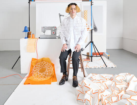
Hearing is Janani Iyer’s passion. As a child, she loved to play music and sing—from classical Indian Carnatic songs to a cappella jazz. During her undergraduate years at the University of California, Berkeley, Iyer studied how the brain processes music and traveled to Boston to work with leading researchers in the field. Before long, her desire to do translational research led her to focus on hearing loss, which afflicts an estimated one in five Americans, according to the Hearing Loss Association of America.
“We don’t have a cure for hearing loss in humans yet,” she says. “One reason for this is that we don’t yet have a way to see inside the inner ear—to visualize and identify biomarkers of hearing loss in humans.”
Today, as a PhD student in the Speech and Hearing Bioscience and Technology graduate program at Harvard’s Graduate School of Arts and Sciences, Iyer combines the life sciences with physics to reveal the hidden structures of the human inner ear. Her goal is to enable clinicians and researchers to see cells and neurons inside the ear so that they can better diagnose hearing loss and develop new ways to treat it.
A Daunting Challenge
Iyer’s love of music followed her to UC Berkeley, where she developed an interest in neuroscience. She didn’t know she could merge these interests, however, until she attended a scientific talk at the Berkley Ear Club, a seminar series hosted by the University’s Department of Psychology. Sitting in a crowd of professors and postdoctoral fellows, Iyer learned about a field of research that investigates the way the brain responds to music—from activity in different regions of the brain to the way that music and language are related, cultural differences in music perception, and much more. She was determined to study music neuroscience.
“When I started college, I was passionate about music and I was eager to study human behavior,” she says. “I never imagined these interests would converge. The Ear Club talk introduced me to a field of research and a community of innovative researchers who were as interested in music as they were in understanding how the brain works. I left feeling confident that I had a future in this field.”
Having found a calling, Iyer faced one major obstacle to answering it: At the time, no faculty at Berkeley were investigating the neural correlates of music perception in humans. So, ahead in her coursework, she came east for seven months to work in the Music and Neuroimaging Lab at Boston’s Beth Israel Deaconess Medical Center. After returning to Berkeley to complete her undergraduate degree, Iyer engaged in a research collaboration with a hearing aid company and was inspired by the translational aspect of this work.
“I realized that I was most motivated and inspired to spend early mornings and late evenings in the lab when I could see how my work would directly impact patients,” she says.

Iyer joined the Molecular Neurotology and Biotechnology Laboratory at Massachusetts Eye and Ear under the direction of Harvard Medical School (HMS) Professor Konstantina Stankovic. There, she shifted her research focus away from the brain and toward the ear, with a focus on developing new imaging technologies that could improve diagnostics for hearing loss.
“If we could physically see which inner ear structures are destroyed over the course of hearing loss in humans, we would have a much better understanding of our therapeutic targets,” she says. “It’s exciting work and I feel privileged to be a contributor.”
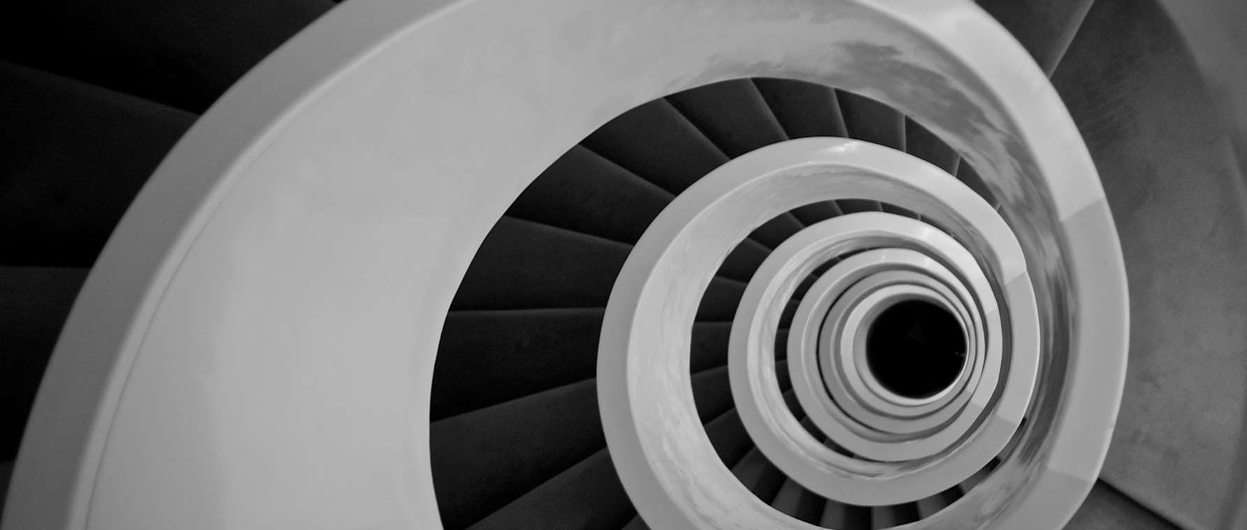
A year after joining Stankovic’s lab, Iyer enrolled as a GSAS PhD student. Since then, she’s remained at the lab, continuing her work with Stankovic and HMS Professor Guillermo Tearney on inner ear imaging. It’s a daunting challenge. To detect biomarkers of hearing loss, scientists need to access the cochlea—the snail-shaped sensory organ that relays acoustic information from the environment to the brain. The structure is small—only about one centimeter across and a few millimeters tall in humans. It’s also deeply encased in the dense temporal bone, which prevents even high-resolution computed tomography (CT) and magnetic resonance imaging (MRI) from accessing its interior.
“The only way we can study human intracochlear anatomy today is by physically removing the bone, slicing the tissue, staining it, and looking at it under a microscope,” Iyer says. “The inner ear cannot be biopsied without destroying hearing.”
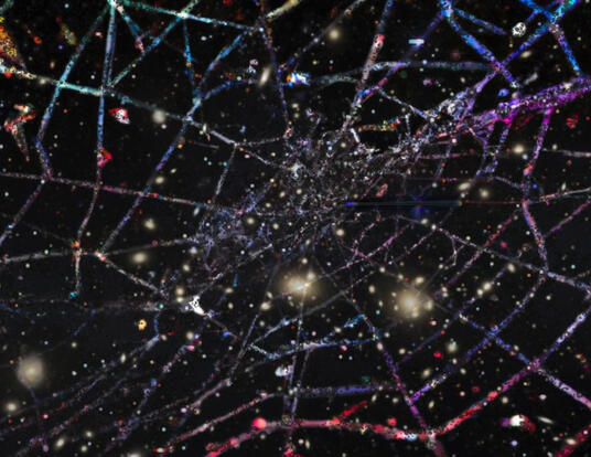
Today, Iyer draws on advanced imaging techniques in her quest to develop non-invasive ways to image the inner ear. Traveling to Canada, she uses a synchrotron particle accelerator to generate powerful X-rays that can penetrate through the aforementioned dense bone surrounding the inner ear, and visually render the tiny structures behind it. The 3D images generated by the process enable scientists to “virtually dissect” tissue with no damage to the body.
“What’s special about this technology,” she says, “is that it leverages high energy X-rays in combination with a phase-contrast mechanism to detect boundaries in a manner than typical X-ray imaging techniques cannot. You can use this powerful X-ray imaging to see the cochlea’s cells and neurons through its dense bony shell, and with additional improvements, this method may serve as a faster and less destructive alternative to the traditional slicing and staining techniques.”
Joining the Fight against COVID
Iyer’s research made her an unlikely recruit in the fight against the COVID-19 pandemic that began last winter. With the illness spreading, though, she wanted to help. She connected with an initiative to manufacture N95 face masks at MIT and eventually was asked to join the MGB Center for COVID Innovation, launched by researchers at Massachusetts General and Brigham and Women’s hospitals.
As co-manager of the Nasopharyngeal Swabs Working Group, Iyer screened and developed testing and clinical validation protocols for 3D-printed and injection-molded nasopharyngeal swabs—ones that would perform similarly to a traditional swab and could also be manufactured quickly at scale. Given the shortage of testing supplies at the time, the work was an important part of the region’s COVID-19 response.
I realized that I was most motivated and inspired to spend early mornings and late evenings in the lab when I could see how my work would directly impact patients.
“The swab has to be able to collect sufficient material while being flexible enough to pass comfortably through the nasal cavity and compatible with testing for the virus,” Iyer says. “We wanted to distribute these swabs throughout the Partners Healthcare Network and also to screen and test technology that will be useful for clinicians throughout the world.”
As supplies have become more plentiful, Iyer has returned to her research on imaging. In addition to her work with the particle accelerator in Canada, she is also developing a miniature inner ear endoscope that resolves the auditory sensory structures using an imaging technique called optical coherence tomography.
“This is a technique that’s already used clinically, for example in ophthalmology and dermatology,” she says. “We know it works and we know it’s safe. So, I’m developing an imaging probe that can be inserted into the cochlea to image the cells and the neurons that we know are critical for hearing in humans.”
Iyer hopes that surgeons who operate on the inner ear will use this imaging tool to improve their technique with the added visual guidance and that researchers will use it to improve diagnostics and therapies for hearing loss. One day, maybe everyone will be able to listen to—and love—music, the way she does.
“Though my research direction no longer has a specific music angle, I am still moved by the fact that the deaf experience music very differently from those who hear,” she says. “To the extent that someone has lost their hearing, for example with aging, and misses the music they enjoyed in their youth, I hope that the information gathered through our inner ear endoscope will help hearing aid and cochlear implant device makers develop improved technologies for music perception.”
Photos courtesy of Jan Iyer, Sergio Fabio Brivio via Creative Commons
Get the Latest Updates
Join Our Newsletter
Subscribe to Colloquy Podcast
Simplecast Stitcher


